Before the invention of microscope, the cell and its existence was not known to man. The study of cell biology has been inseparable from the evolution of the microscope. The 19th century was an era in which microscope contributed a lot to the extensive study of cell. The limit of resolution of an unaided human eye is about 100 microns under optimal viewing condition. Thus, our eyes cannot see those objects distinctly which are separated by a distance less than 100 microns and the object itself smaller than 100 microns. The limitation of human eye helped in the evolution of various magnifying instruments. First microscope was a simple magnifying glass (convex lens) then compound microscope. In modern times electron microscope gradually extended the resolving power 1,00,000 times greater than those of the optical microscope.
Microscopy
Microscopes have been important optical instruments in the study of cells and other minute objects because they possess high magnification and high resolving power to distinguish between two objects lying very close together. The lesser the distance that can be measured between two objects in a microscope the greater is its resolving power. The capacity of the microscope, both to magnify and resolve, depends on the kind of illumination used i.e. the wavelength of light. Because of the nature of light, microscope using light as a source of illumination cannot resolve objects less than one-half the wave-length of the light.
The average wave-length of white light is about 0.55 microns. Therefore the microscope using white light cannot resolve objects less than about 0.2 microns. Compound microscope is used to produce a considerable magnification of small objects. The essential part of the instrument consists of two convex lens systems, placed co-axialy within two sliding tubes at some distance from each other. The first lens which remains towards the objects is called the objective lens. It is of very short focal length and of small aperture.

The second convex lens of microscope through which observations are made is called the ocular lens or eyepiece. It is of larger focal length and of bigger aperture than the objective lens. The objective lens produces an initial image of the object. The eye lens magnifies the initial image and produces the final image. To see objects smaller than 0.2 microns, a electron microscope is used where a beam of electrons is used as a source of illumination. They have the wave length of about 0.50 Å. Thus, the resolving power of the electron microscope can be one half of the 0.50 Å. The electron microscope was developed in 1940s. It was based on matter waves associated with electrons.

Electron microscopy has made a considerable increase in resolving power of the instrument. The electron microscope since the time of its design has proved a powerful tool of great value for both science and industry. The major difference between the electron microscope and the light microscope, is the physical design of the two instruments. The electrons can travel to a good distance in vacuum so the entire electron microscope unit is enclosed in a vacuum tight chamber.

A narrow beam of electrons is emitted by a metal filament heated in a vacuum column. The electron beam is collected and focused upon the specimen by the electro-magnetic condenser lens. Electrons are collected by the electro-magnetic objective lens after passing through the object.
The objective lens enlarges the image of very minute objects. The electro-magnetic projector or ‘eyepiece’ lens further magnifies the image of the object and projects it on a flouroscent screen or on the photographic plate.
Phase Contrast Microscope : This microscope is used for the study of living cells which are, in general, transparent to light. The principle on which this microscope is based is that the light passing through an object undergoes a phase retardation which normally is not detected. In this instrument, however, the phase difference is changed to one-fourth of the wavelength and the small variations in phase produced by the various structures are thereby made visible.
Ultraviolet Microscope : The ultra violet microscope looks like a compound microscope. It differes from the light microscopes in its lenses. The lenses are made up of quartz which can transmit ultra violet light. The ultra violet microscope is very much helpful in the study of chromosomes and nucleic acids (DNA and RNA) because they absorb ultra violet light then cytoplasm.
Polarising Microscope : The polarising microscope resembles a compound microscope. The polarising microscope can detect regions in cells where constituents are disposed in highly ordered array. This is done with the help of a prism. The prism transmits plane polarized light instead of ordinary light.
Dark Field Microscope : The dark field microscope is an ordinary microscope with a special condenser. This condenser stops the beam of light from the centre of field and the object is illuminated only by oblique beam of light. Due to oblique beam of light, this microscope is suitable for the study of outlines of cells, nucleus, oil droplets vacuoles, and mitochodria etc.
Cytochemistry : Cytochemistry is the branch of science which deals with the study of chemical organisation by using special methods of preparation. The chemical organization is studied by producing a colour contrast. To produce a colour contrast various dyes are used. For example, to localise DNA, Feulger dye is used under certain condition; similarly to localise both RNA and DNA Methyl green pyroism is used. It is not possible for us to localize a large number of cell constituents by the method of cytochemistry.
Autoradiography : Autoradiography is a well known and most important modern cytochemical methods to study the synthesis of molecules and to trace the metabolic events in the cells. This is done by use of substances labelled with radio isotopes. The isotopes which are mainly used are tritium (3H), carbon (14c) and phosphorus (32P). Tritium, a carbon labelled thymidine, is used for studying the synthesis of DNA; tritium or carbon labelled uridne is used for studying the synthesis of RNA; and tritium or carbon labelled amino acids are used for tracing proteinsynthesis.
Cell Fractionations : This involves the isolation of pure fractions of cell like nuclei, mitochondria, chloroplast, golgi bodies, ribosomes, lysosomes etc. The phenomenon of centrifugation has played a important role in cell fractionation. The cells or tissues are ground up and homogenized in sucrose solution and then are centrifuged. The successive centrifugation of the resulting homogenate produces fraction depending upon their weight and size. This method of separation of cellular components by centrifugation is called the differential centrifugation.
Biochemical Techniques : Widely used bio-chemical methods are given below in brief :
(a) For biochemical analysis a pH meter is used to measure accurately the pH of a solution.
(b) Chromotagraphy is another important biochemical technique to separate small quantities of organic (and inorganic) compounds depending upon the rate of their flow on the chromatographic paper.

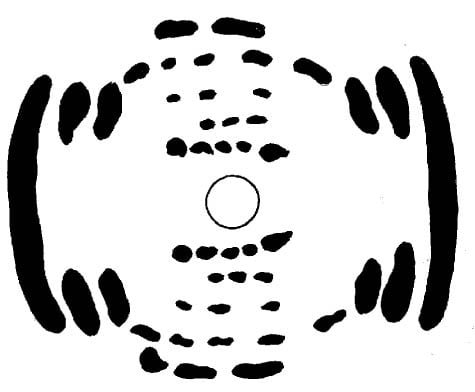
(c) X-ray crystallography is used to determine the molecular organization of substances. This technique was used for finding the molecular configuration of double helix of DNA by Waston and Crick.
Tissue Culture : In this technique isolated cells are cultivated in special fluid media in varieties of glass plastic tubes, vials or bottles.

