
Circulatory System : It is a closed system of heart, arteries and veins which are used for transporting the blood to all parts of the body.
Blood: It is a liquid connective tissue present in blood vessels inside the body. In its plasma the blood corpuscles or cells are floating.
Plasma : It is a yellow straw coloured fluid in which blood corpuscles are not present. It consists of 90-92% water and 10-8% solids.
Blood Cells
(i) Red Blood Corpuscles or Erythrocytes (RBC) : These are the red cells which are cicular disc shaped cells having no nucleus. They contain haemoglobin (an iron containing pigment). They act as carriers of CO2 and O2 . In a man there are 5-5.5 million RBC per mm3 of blood and in woman it is 4.5-5 million per mm3.
(ii) White Blood Corpuscles (WBC): These are bigger in size than RBC and are colourless due to absence of haemoglobin. They have a nucleus. Their number is between 6000-10000 per mm3 of blood.
(iii) Blood Platelets : They are called thrombocytes. They have very small irregular structure. Their number is about 250000 per mm3 of blood.
They play an important role in coagulation of blood or clotting of blood.
Heparin : It is an anticoagulant substance present in blood that prevents it from clotting.
Thrombosis : It refers to the intravascular clotting.
Hirudin : It is an anticoagulant (found in leech).
Types of circulation Sysytem
(1) Intra-cellular circulation : Circulation within single cell like amoeba.
(2) Extra-cellular circulation : Circulation outside the cell, sub divided as follows:
(a) Water vascular system : For example sponges, coelenterates, echinodermates. In marine animals water enters the body cavities.
(b) Intra-organismic circulation : In certain higher non-chordates including annelids and in all chordates, this type of circulation is present. These organisms need more supply of oxygen which is supplied quickly by circulatory system present in them. This type of circulatory system can be divided into the following two types.
(i) Blood vascular system : This system is present in higher nonchordates (including annelids) and chordates. It consists of blood vessels, blood sinuses and heart.
(ii) Lymphatic system : Lymphatic system or Blood vascular system can be divided into following two types :
Blood circulatory System
(1) Open type of blood circulatory system : The circulating fluid (known as haemolymph or perilymph) is not only present in the vessels but also present in the blood spaces or lacunae or sinuses e.g. some arthropods, molluscus and annelids.
(2) Close type of blood circulatory system : It is present in most of the annelids and all vertebrates including man.
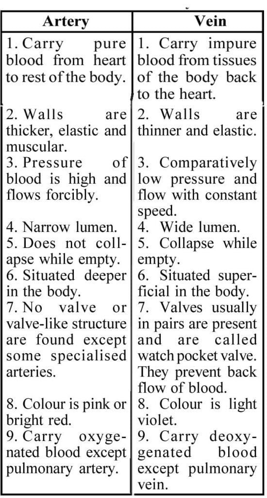
Blood Vessels
The blood remains confined in the blood vessels (arteries and veins)
Heart
Heart is a voluntary muscle and a highly modified blood vessel which has the capacity to contract and expand throughout the life. It pumps the pure blood into the arteries and receives the impure blood through the veins. It has three layers present in it as follows :
(1) Epicardium : Similar to tunica externa.
(2) Mesocardium : Similar to tunica media.
(3) Endocardium : Similar to tunica interna.
Generally heart can be of the following types :
(i) Venous heart : When only impure blood (i.e. venous blood) is pumped by the heart, e.g. fishes.
(ii) Mixed heart : In the heart of amphibians and most of the reptiles, the oxygenated and deoxygenated blood is mixed. Such type of heart is a mixed heart.
(iii) Double circulatory heart : In the heart of crocodile (reptilia), birds and mammals, oxygenated and deoxygenated blood do not mix in any part of auricle and ventricle. There is a separation of blood circulation completely in the heart. The left half of the heart contains oxygenated and right half contains deoxygenated blood. Thus there is a double circulation in the heart.
Vasa vasorum : Vasa vasorum are the blood vessels supplying blood to the walls of blood vessels.
Working of Heart
(1) A normal human heart beats approximately 72 times in a minute.
(2) There are two centres of contraction present in the heart of man. One in the right auricle known as sinuauricular node (SA node). It is also known as pacemaker. The other is present at the junction of right and left ventricle known as atrio-ventricular node (AV node).
(3) Both of these nodes are modified cardiac muscles and supplied with the parasympathetic and sympathetic nerves.
(4) Working of heart is myogenic.
(5) When heart beat starts, the auricles and the ventricles are in relaxed form. This stage is known as joint diastole. In this stage blood comes in the auricle.
(6) Then comes the systolic phase. In this phase contraction of auricle takes place which forces pumping of blood into their respective ventricle.
(7) The above action is due to the excitation of the SA node which sends the waves of contraction. These waves contract the right auricle first then they go towards the left auricle and contract.
(8) After the contraction of the left auricle the waves of contraction are centralised at the AV node.
(9) This causes the stimulation of AV node and this node sends the electrical stimulation to the apex of ventricle via ‘bundle of His’.
(10) When this stimulation reaches the apex of ventricle, this is given to the Purkinje fibres present in the wall of ventricle. From its apex the contraction reaches towards the anterior side and thus blood is pumped into the aorta or arches originating from ventricle.
(11) After the systolic phase the diastolic phase starts.
Sound of murmur : Some disorders of heart value create sounds of murmur. It may be due to—
1. Valvular insufficiency
2. Stenosis
Sounds of heart : The sounds are produced due to the closure of valves of heart.
First sound : It is due to the closure of atrioventricular valves. This sound is like LUBB.
Second sound : It is due to the sudden closure of semilunar valves present at the opening (origin) of aorta or arches from the ventricles. This sound is like DUP.
Thus sound of heart is LUBB-DUP and a pause and again LUBB-DUP will develop.
Blood pressure : The pressure by which blood flows in the arteries is called blood pressure. It is measured by sphygmomanometer.
Normal systolic pressure = 125-130 mm Hg
Normal diastolic pressure = 70-90 mm Hg
Electrocardiogram : It is an instrument which measures different stages of potential in the form of a graph known as E.C.G. A typical E.C.G. has—
1. P-wave : Depolarization of auricle.
2. Q.R.S. Complex : Depolarization of ventricle.
3. T-wave : Repolarization of ventricle.
Cardiac output : This is the volume of blood pumped by the heart in a minute.
C.O.=Heart rate × Stroke volume
Blood Coagulation
Blood coagulation is carried out by change of fibrinogen into fibrin. Fibrin is insoluble fibrous protein.
Following steps are involved in blood coagulation.
(1) Liberation of thromboplastin : Thromboplastic activity starts at the time of clotting by some factors viz. Ca++ ion, antihaemophilic factor (AHF) and phospholipid.
(2) Conversion of prothrombin into thrombin : Ca++ ion converts prothrombin into thrombin or accelerates globulin.
(3) Conversion of fibrinogen into fibrin : This conversion is carried out by thrombin in the presence of factor F-XIII.
(4) Action of blood platelets : It is well known that thromboplastin is present in the platelets which are broken down and liberates thromboplastin at the time of injury.
Factors responsible for clotting
Fibrinogen
Prothrombin
Thromboplastin
Calcium
Labile factor or proaccelerin
Accelerin (Hypothetical factor)
Stable factor or autoproth rombin (From serum)
Antihaemophilic factor (AHF)
Christmas Factor
Stuart power factor (From serum)
Plasma thromboplastin antecedent (PTA)
Hageman factor or contact factor
Fibrin stabilizing factor or fibrinase
Bleeding time : It is the time taken till the bleeding continues after a given incision. It is about 3-6 minutes.
Clotting time : It is the time taken in clotting the blood after giving a incision and coming out of blood. It is about 5-8 minutes.
Lymphatic System : This system has,
1. Lymph
2. Lymph vessels
3. Lymph glands
Lymph : Lymph is like blood without RBC. It has the property of clotting when it comes in contact with air.
During the blood circulation plasma and WBC comes out of the blood capillaries and enters in the lymph capillaries, it is known as lymph. This lymph is again sent to the blood circulation. The blood and lymph circulation continues.
Lymph vessels : These are thin vessels. The smallest vessels are called capillaries which unite to make lymph vessels. These vessels are provided with valves which do not allow the backflow of lymph in the vessels.
Only two main lymph vessels are present in the man.
1. Thoracic duct : It is present in the wall of thorax and abdomen. This duct opens into left subclavian vein.
2. Right thoracic duct : Lymph vessels from right side opens in it. It opens into right subclavian vein.
Lymph glands : Spleen and lymph nodes are called lymph glands.
Functions of lymph
1. It forms the lymphocytes.
2. The glands act as centre for the formation of antibodies.
3. It absorbs the fatty acids and glycerols from the alimentary canal and transports it to the different parts of the body.
4. Lymph transports nutrients from blood to the tissue cells.
Allergy : The allergies include a group of non-infectious reactions which occur due to the hypersesitiveness of certain individuals to foreign matter that may enter the body or come in contact with body. Dust, fibers, feathers, pollen etc. may act as allergens.
Allergens are the foreign substances that cause allergy. They are also known as antigens.
Antibodies : Antibody is a plasma protein which belongs to the group called gamma globulin.
Thrombosis : The coagulation of blood within the blood vessel or heart during lift is called thrombosis.
Serum : It is the liquid which forms when blood clots.