
Introduction : As bricks are units of any building, cells are the building blocks of any plant or animal body. All living organisms are made up of cells.
The body of a plant or animal may be unicellular or may be made up of many cells (multicellular). In a single cell organism the characteristic features of life are expressed by the single cell. In multicellular organisms, all the functions of life are carried out by group of cells.
Schleiden and Schwann were the first to tell that plants and animals are made up of cells.
They proposed cell theory in 1838.
Cell Theory : Cell theory may be described as follows :
1. All plants and animals are composed of cells.
2. Cell is the functional unit of every living being.
3. All cell arise from pre-existing cells.
4. All cells are basically the same.
In short, we can say that basic unit of structure and function in living organisms is the cell.
The cell has been defined in different ways by different scientists.
(i) According to Loewy and Siekevitz 1969, the cell is a unit of biological activity and is capable of self reproduction.
(ii) According to Swanson and Webster 1978, cell is a physical entity and is the basic unit of life.
(iii) According to De Robertis 1981, the cell is the smallest unit of life.
According to some discoveries—
(a) Van Mohal and Nageli in 1849 declared independently that the two main parts of the cell are the cell wall and the cell contents.
(b) Colin in 1850 proved experimentally that the protoplasm of plants and animals are identical.
(c) Robert Brown discovered the nucleus of of the cell in 1831.
Size of the Cell
Size of the cells varies widely in different organisms. The size of the cells is measured in Angstrom (Å), millimicron (mm) and micron (m). Smallests cell of 0.1m is of pleuro pneumonia and the largest is Ostrich egg asbout 16 cm across. Man’s brain’s nerve cells are the largest—about one meter in length. In plants, fibres of Jute, and Cotton, etc. have the largest cells.
Shapes of Cells
Some cells are flat (skin), elongated (muscle) Columnar, elongated, disc shaped, oval, etc.
Types of Cells
Cell are of two types—
(a) Prokaryotic cells.
(b) Eukaryotic cells.
(a) Prokaryotic Cells : These are the most primitive cells. They lack true nucleus. Examples of these cells are—Bacteria, blue-green algae, etc.
(b) Eukaryotic Cells : These are the cells with true nucleus. Nucleus is always enclosed by a nuclear membrane.
These cells are present in plants as well as in animals.
Parts of the Cell
(i) Cell wall (only in Plant cells)
(ii) Protoplasm
Cell membrane or
Plasma membrane Nucleus Cytoplasm
Nucleus Nuclear membrane
Nucleoplasm
Nuclear reticulum
Nucleolus
Cytoplasm Cytoplasmic matrix
Cytoplasmic organelles
Cytoplasmic organelles mitochondria
Centrosome (only in animal cells)
Golgibody
Plastids (only in plant cells)
Lysosomes
Endoplasmic reticulum
Cillia and flagella
Plastids Chloroplasts
Chromoplast
Leucoplasts
Description of these parts is given below—
Cell Wall : (Cell wall is found only in plant cells) A plant cell has two parts—cell wall and protoplast. Protoplast is a living substance. It is bound by plasma membrane. Cell wall is outer, rigid porous, non-living covering of cells. It is made up of cellulose. Cellulose is Carbohydrate. Cell wall also contains pectin, lignin, etc. Cell walls are two—primary and secondary cell wall.
The cell wall gives shape and rigidity to the cell and protects protoplasm. It contains water. Cell walls also contain pits.
Protoplasm : The transparent, colourless, colloidal fluid as a living substance is called protoplasm. It contains numerous suspended granules and droplets. It contains about 85% water, 10% proteins, 1.5% fats, 1% carbohydrates.
Cell membrane or plasma membrane : It is a thin flexible semipermeable living membrane. It surrounds the protoplasm of a cell. Cell membrane is found in all plants and animal cells. Membranes of different organisms are different in thickness and function. The thickness is about 20 to 30 Å. These membranes are visible with an electron microscope.
Cytoplasm : The part of the protoplasm that surrounds the nucleus is called cytoplasm. On its outer side is plasma membrane. Tonoplast is on the inner side. Cytoplasm is a complex material composed of membranes, particles and organelles.
The cytosol is the soluble part of the cytoplasm. The fluid part of the cytoplasm is called hyaloplasm. The fluid filled cavities are called vacuoles.
Cytoplasm supplies proper nutrition to the living parts of the cell. Many vital and chemical reactions important for life take place in it. These reactions are important for protein synthesis, respiration, etc.
Nucleus : It is spherical, dense and centrally located portion of the cell. It occupies two thirds of the cell space. It contains hereditary material i.e. chromosomes. Some cells may contain more than one nucleus.
A nucleus consists of the following four parts, namely:
(i) Nuclear Membrane : The membrane which separates the nucleus from cytoplasm is known as nuclear membrane. In the nuclear membrane many pores can be observed.
(ii) Nucleoplasm : Nucleoplasm is a dense clear mass of protoplasm. It is filled in the nucleus. It contains materials used in cell division. The chromosomes and nucleolus are embedded in the nucleoplasm.
(iii) Nuclear Reticulum : Thread like material embedded in the nucloplasm is called nuclear reticulum. The threads of this network are called chromatin. Chromosomes get developed from reticulum.
(iv) Nucleolus : The round dark stained acidophilic body situated in the matrix of nucleus is called nucleolus. Nucleolus involves in the formation of ribosomal sub-units.
Nucleus is the control centre of the cell. It contains the hereditary material in the chromosomes.
Plastids : Plastids are granular cell organelles. Plants use them for manufacture and storage of food materials.
Plastids are present in plant cells. Their number is different in different plants. Plastids are of three types—(a) Chloroplast, (b) Chromoplast and (c) Leucoplast.
(a) Chloroplast : These are green chlorophyll pigments. These mainly are found in green plants’ parts such as leaves. These are oval or discoid in shape. Chloroplasts trap solar energy and change it into chemical energy by the process of photosynthesis.
(b) Chromoplast : These are also coloured but their colour is not green. These are found in the petals of flowers, skins of fruits etc. In shape, chromoplasts are elliptical in shape. Usually these contain orange pigments. Bright colours of petals are due to chromoplasts.
(c) Leucoplasts : These are colourless plastids. Leucoplasts are mainly found in underground roots. These are not exposed to sun light. In size they are small and in shape they are spherical. Leucoplasts transform sugar into starch. The food storage is done by leucoplasts.
Mitochondria : Mitochondria is mainly responsible for the release of energy from food material. Mitochondria is found in the cells of all the plants and animals. Mitochondria is not found in bacteria and blue-green algae. The size of mitochondria varies from 0.2 to 7.0m in diameter.
The mitochondria are considered as ‘power house of the cell’. They are the aerobic respiratory centres of the cell. They are essentially energy converters.
Golgi Body : As the name indicates, golgi body or golgi apparatus was discovered by Camillo Golgi in 1898. They occur near the nucleus. Golgi appartus are present in all animal and plants’ cells. They occur as network of threads. They are involved in secretory activity of the cell.
Lysosomes : These are subcellular particles of cytoplasm. They have sac-like structure. Mainly they occur in animal cells. Lysosomes are membrane bound vesicles. They are spherical in shape. Lysosomes contain some digestive enzymes which help in the digestion of food. Lysosomes also provide defence against bacteria and viruses.
Endoplasmic Reticulum : It is a system of folded membrane in the cytoplasm. It does the work of protein synthesis. These are found in all animals and eukaryotic plants. They are found near the nuclear membrane.
It is a very complex network of double membranes. These are responsible for many biochemical reactions. Endoplasmic reticulum also provides mechanical support to colloidal cytoplasm.
Ribosomes : Ribosomes are minute granular structures present in the cell. They are of spherical shape. Ribosomes are present both in plant and animal cells. Their main function is the synthesis of protein in the cell.
Cytoskeleton : Cytoskeleton maintains the shape of the cell. It contains protein filaments. Cytoskeleton regulates distribution and orientation of organelles.
Centrioles and Centrosome : Centrioles are hollow cylinders and do not have membranes. They occur in pairs at right angles to one another. Centrioles are found in most of the animal cells and lower plant cells. It is made of nine triplets of microtubules. They separate and migrate to opposite poles of an animal cell. Cilia and flagella development and organisation is also done by Centrioles.
Centrosome is round in structure in the cytoplasm near the nucleus.
Cilia and Flagella : Cilium is hair like protoplasmic process which extends from the cell surface. Whip like protoplasmic extensions of the cell surface is known as flagellum. Plural of cilium is cilia and that of flagellum is flagella.
These are present both in eukaryotic and prokaryotic cells. From structural point they contain nine pairs of peripheral tubules. Cilia and flagella beat rhythmically and propel a cell through liquid medium.
Non-Living Objects
Vacuoles : Vacuoles are liquid filled areas of the cell surrounded by unit membrane. Vacuoles are found both in plants and animal cells. Their number, size and position in different cells are different. Their size is small. They contain dissolved material like sugar, salts, organic acids etc. They act like reservoirs of various secretory and excretory materials. They also provide water storage space within the cells.
Ergastic Substances : Ergastic substances are the non-living waste substances which are present in the cell sap. These substances are of three types—
(a) Stored matters.
(b) Secreted substances.
(c) Alkaloids.
They control physiological processes in plants. They are also used as economic products.
Chromosomes
Chromosomes control the activities of a cell. Chromosomes mainly transfer of genetic traits from one generation to the next and hence referred to as ‘Physical basis of heredity’.
Chromosomes are long, cylindrical structures and appear during cell division. Plant chromosomes are bigger in size than animal chromosomes.
The chromosomes are stout and rod shaped during metaphase but appear in the shapes of V, L, J, I during anaphase of cell division.
In the nuclei of plant and animal cells, the number of chromosomes is represented by diploid condition which shows two sets of chromosomes. Vegetative cells generally show diploid condition (2n). In gametes, only one set of chromosomes is found. This is called haploid condition (n).
Karyotype : A single set of chromosomes present in an organism is called ‘karyotype’.
Idiogram : The diagrammatic representation of a single set of chromosomes is called ‘idiogram’.
Genome : The genes constituting a single set is called ‘genome’.
Alleles : Every character is determined by a pair of genes called ‘alleles’.
Centromere : The metaphase chromosome shows a colourless constriction called ‘centromere or kinetochore of kinomere’. The centromere helps in the attachment of spindle fibres.
Chromatids : On both the sides of the centromere there are long cylindrical parts called arms which longitudinally splits into two parts called chromatids. The two chromatids are spirally coiled around one another.
Telomeres : The ends of chromatid arms exhibit polarity and are called ‘telomeres’.
Chromomeres : During early prophase the chromosomes appear as beads with swellings and constrictions alternating with one another. The swellings are known as ‘chromomeres’.
Satellite or Trebent : The part of chromosome arm present beyond the secondary constriction is called a ‘satellite or trebent’.
Nucleolar organisers : Some chromosomes have secondary constrictions from which nucleoli are formed. These are known as ‘nucleolar organisers’.
Chemical nature chromosomes: Chromosomes are composed of nucleo-proteins.
Heterochromatin : When chromosomes are stained with acetocarmine or acetoorcein, some parts take more stain and appear darker. These areas are called ‘heterochromatin’.
Euchromatin : Some parts of chromosome take less stain and appear lighter in colour. These areas are called ‘euchromatin’.
Classification of chromosomes
Based on the number of centromeres.
Acentric centromere is absent
Monocentric one centro mere is present
Dicentric two centromeres are present
Holocentric number of centrol- meres is not definite.
Polycentric many centromeres are present.
Based on the Position of the Centromere
Metacentric : Centromere is present in the middle point. Both arms are equal in length. During anaphase they appear in V shape.
Submetacentric : Centromere is slightly away from the middle point and arms are unequal. During anaphase they appear in L-shape.
Acrocentric : Centromere is present one side on which one arm is very long and the other arm is very short. During anaphase, they look in J-shape.
Telocentric : As centromere is present at the end of the arm, only one arm is present. They appear in I-shape.
Autosomes : Autosomes determine the vegetative activities of cell like growth, development, etc. They are more in number.
Allosomes : Allosomes are called as sex chromosomes. In human beings they are identified as X, Y chromosomes.
Polytene chromosomes : These are giant chromosomes, found in the salivery gland cells of Drosophila belonging to the order Diptera.
Lamp brush chromosomes : These are giant chromosomes occurring in the oocytes of amphibians. They show a central axis on which large number of loops are formed which look like bristles of a brush.
Nucleic Acids
The nucleic acids are giant molecules present in the nucleus. They carry a number of biochemical activities of cell. It leads to the growth and development of organisms.
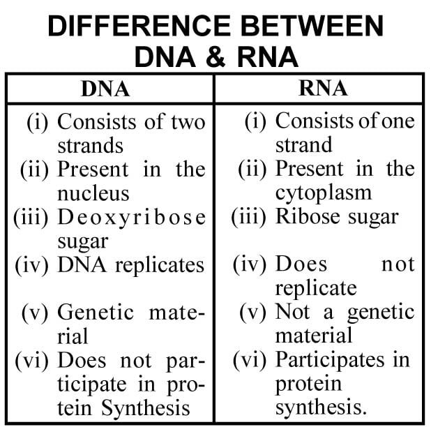
DNA
Deoxyribo Nucleic Acid (DNA): DNA is mainly concentrated in the chromosomes and is the chief genetic material in all organisms except in plant viruses. Hershey and Chase (1954) confirm DNA as the generic material. It is refered as the chemical basis of heredity.
Structure of DNA : The structure of DNA was proposed by J.D. Watson and F.H.C. Crick in 1953. Watson and Crick’s model is known as ‘double helix’. It is composed of two strands which are spirally coiled around one another. DNA looks like a twisted ladder.
In DNA double helix, the length of each coil is 34 Å and contains 10 equally spaced base pairs. The distance between two successive base pairs in 3.4 Å and the angle between them is 36O.
Properties of DNA
1. It is a double helix.
2. It has a very high molecular weight.
3. It shows high absorption spectrum in ultraviolet region.
4. It is denatured when heated upto 70OC.
5. High pH and low salt conditions also cause denaturation of DNA.
6. DNA is the chief genetic material of all organisms except plant viruses.
DNA Types : B-DNA is more common and has clockwise helix structure. It shows a definite hereditary stability.
Z-DNA is uncommon and has an alternating clockwise and anticlockwise helix structure.
Functions of DNA
Autocatalysis or Replication : The process of duplication of a single DNA molecule into two daughter DNA molecules is called ‘autocatalysis’.
Heterocatalysis : DNA promotes the synthesis of proteins and regulates various biochemical reactions of cell.
RNA
Ribose Nucleic Acid (RNA) : It is mainly concentrated in ribosomes and also found in plastids, and mitochondria in traces. It is synthesized inside the nucleus with the help of the enzyme called ‘RNA polymerase’, but is later released into cytoplasm.
RNA does an important work in protein synthesis and is non-genetic.
Structure of RNA : RNA is made up of a single polynucleotide strand. The nucleotides of RNA comprise of three components called phosphate group, ribose sugar (C5H10O5) and nitrogen bases.
Types of RNA
Messenger RNA : It is a unfolded polynucleotide molecule consisting of about few hundred nucleotides. It is synthesized from DNA template chain by a process called ‘transcription’.
2. Ribosomal RNA : It is obtained in the ribosomes and constitutes about 75-80% of the total cellular RNA.
Transfer RNA : It is called ‘soluble RNA or adaptor RNA’. It constitutes about 15% of the total RNA.
Cell Division
All new cells come out from pre-existing cells by cell division. This theory was proposed by Rudolph Virchow in 1859.
The cell division is of three types : (a) Amitosis (b) Mitosis and
(c) Meiosis.
(a) Amitosis : Amitosis is a simple type and direct cell division. During the division the nucleus elongates and becomes dumb-bell shaped. It finally splits into two daughter nuclei. The splitting of nucleod is followed by constriction of cytoplasm. The cell is divided into two equal halves. It mainly occurs in bacteria yeast and Amoeba cells.
(b) Mitosis or Equational Division : It occurs in vegetative cells. In this division the mother cell produces two daughter cells. These two cells are identical in shape, size and character. That is why this is called equational division. The cell number increases by mitosis by which the growth of the organism also increases.
Cell cycle and stage of cell cycle: The sequence of events from the formation of cell and upto the division of the cell into two daughter cells is called cell cycle.
Cell cycle has two stages—
(A) Interphase and (B) Mitotic phase.
(A) Interphase : The period between two successive divisions of the cell is known as interphase. It is the most active period for reparing cell for division.
The interphase is sub-divided into three distinct stages, called—(i) G1 phase (ii) S-phase and (iii) G2 phase. G-stands for gap period and S-stands for synthesis.
1. G1 phase : In this phase the size of the cell increases and a lot of RNA and protein are synthesised.
2. S-phase : In S-phase, the DNA present in each chromosome gets duplicated. This is followed by the splitting of chromosome arm into two chromatids which are united at centromere.
3. G2 phase : In G2 phase the synthesis of protein and RNA continues.
(B) Mitotic phase : At the end of interphase, the cell is ready for division. In this phase the nucleus divides first and it is followed by division of cytoplasm. Nuclear division is called ‘karyokinesis’ and cytoplasmic division is called ‘cytokinesis’.
(a) Karyokinesis : In this the nucleus undergoes a number of sequential changes. It is sub-divided into four phases called Prophase, Metaphase, Anaphase and Telophase.
(1) Prophase : Prophase is the first stage of nuclear division in which the prochromosomes are long, slender and they spread extensively in the nucleoplasm. They gradually become short, thick, stout and develop into rod shaped chromosomes by ‘coiling’ process.
In the last stage of prophase, the nuclear membrane dissolves and the nucleolus decreases in size and finally disappears. The chromosomes appear to be randomly scattered in the cytoplasm.
(2) Metaphase : A bipolar apparatus is formed in metaphase by the fusion of microtubules.
(3) Anaphase : In anaphase the spindle fibres begin to contract causing pressure on the centromeres. As a result the centromere of each chromosome divides and thus, the two chromatids are separated.
(4) Telophase : The daughter chromosomes arrived at the poles become thin, long, and lose their visibility due to despiralisation to form chromatin. The nuclear membrane reappears and the nucleoli are reorganised from their relevant chromosomes. In the final stage of telophase, two independent daughter nuclei are organised.
(b) Cytokinesis : The cytokinesis is marked by the formation of cell wall in the region of equatorial plate. At the end of the telophase, soon after the formation of daughter nuclei, the spindle fragments gather at the equator region and form a barrel shaped structure, called ‘phragmoplast’.
Significance of Mitosis
1. Mitosis causes the organism growth.
2. The daughter cells formed in mitosis are identical with the mother cell.
3. In unicellular species, mitosis helps in reproduction.
4. It is useful in the wear and tear mechanism of the plant body and is also useful in the healing of wounds.
5. It is useful in the regeneration of lost parts, and for grafting in vegetative reproduction.
Stages of Meiotic Cell Division
Meiosis takes places in two stages, called—(1) Meiosis-I, and
(2) Meiosis-II.
Meiosis-I is heterotypic division and in this the diploid parent nucleus divides into two daughter nuclei each having haploid set of chromosomes. Meiosis-II is homeotypic division in which two haploid nuclei divide mitotically and form four haploid.
(c) Meiosis : Meiosis takes place in the germinal cells. In this division, the double number of Chromosomes (diploid) is reduced to its half number (haploid). This is called meiosis and gametes are unidentical with that of the mother cell. That is why it is called, ‘heterotypic division’.
Stages of Meotic Division
There are two stages of meotic division namely Meosis-I and Meosis-II.
Meosis-I : It is heterotypic division. In this diploid parent nucleus divides in to two daughter nuclei each having haploid set of chromosomes.
Meosis-I has four phases namely—
1. Prophase-I
2. Metaphase-I
3. Anaphase-I
4. Telophase-I
Prophase-I further has five substages, called—(a) Leptodone
(b) Zygotene (c) Pachytene
(d) Diplotene (e) Diakinesis.
Meosis II : It is homeotypic division. In this division two haploid nuclei divide mitotically and form four haploid cells.
Meosis II also has four phases as—
(a) Prophase-II
(b) Metaphase-II
(c) Anaphase-II
(d) Telophase-II
Cytokinesis : After each nuclear division cytoplasm division can occur. The process of cytokinesis is same as that of mitotic division.
Significance of Meosis
(i) Constant chromosome number is maintained from one generation to the next by meosis.
(ii) Formation of gametes takes place in meosis; and that is why it is useful in sexual reproduction.
(iii) New species are originated by meosis and thus it leads to evolution—
