The cell is the basic unit of life.
Cell : Cell is the basic unit of structure and function in living organisms.
Cell Theory
1. All living things are composed of cells.
2. All cells arise from pre-existing cells.
3. All cells in chemical composition and metabolic activities are alike.
4. The function of an organism depends totally on the activities and interaction of the constituent cells.
Types of cells : Typically cells are of two types depending on the type of nucleus.

These are : (a) prokaryotic cell and (b) eukaryotic cell.
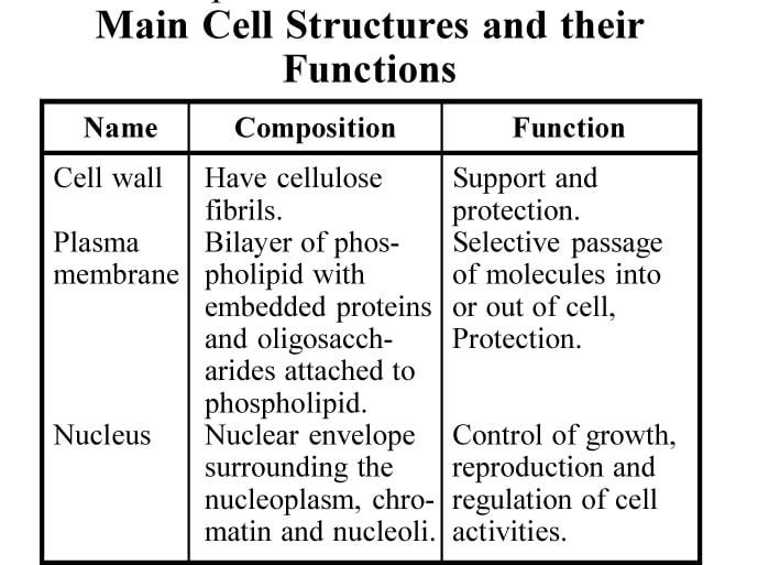
Cell wall : Cell wall is the outer, rigid, porous, non-living covering of plant cells. It is found only in plant cells.
Primary cell wall : Primary cell wall is the external part of the cell. It is thinner than the secondary cell wall.
Secondary cell wall : Secondary cell wall is formed after the formation of primary cell wall. It is the inner part of the cell wall. It is much thicker than the primary cell wall.
Middle lamella : The middle lamella is the original wall between two adjacent cells, on which the thickening layers have been deposited.
Plasmodesmata : Plasmodesmata are the very small pores in the cell wall through which delicate threads of cytoplasm pass and cytoplasmic connections between two adjacent cells are established. Each one of the fine protoplasmic fibres is called plasmodesmata.
Plasma Membrane : The protoplasm of a cell is surrounded by a thin flexible, semipermeable living membrane. This is called plasma membrane or plasmalemma. Cell membrane is present in all plant and animal cells.
Functions of cell membrane : It has three functions viz. (a) Cell membrane protects the protoplasm and other cell organelles from external damages.
(b) Provides mechanical support to animal cells. (c) It acts as a controlling device to molecules.
Endocytosis and exocytosis : Endocytosis and exocytosis are involved in the bulk transport of materials through membranes, either into cells (endocytosis) or out of cells (exocytosis).
Phagocytosis : In cell eating materials are taken up in solid form. Cells specialising in the process are called phagocytes and are said to be phagocytic; e.g., some white blood cells of blood.
Pinocytosis : In this materials are taken up in liquid form. Vesicles formed are very small, in which case the process is known as micropinocytosis and the vesicles are known as micropinocytoic.

Cytoplasm : The cytoplasm surrounds the nucleus of a cell and is the viscous, homogeneous, granular living material. It is a complex material made up of membranes, particles and several organelles excluding nucleus.
In all living plants and animal cells cytoplasm is present. In a cell it occurs in the region extending from plasma membrane of nuclear membrane.
Hyaloplasm or cytoplasmic matrix : Hyaloplasm or cytoplasmic matrix is the fluid part of the cytoplasm.
Vacuoles : The vacuoles are the fluid-filled cavities.
Cell sap : Cell sap is the fluid present in the vacuole. It contains dissolved materials.
Ectoplasm : Ectoplasm is the outer non-granular, transparent thin layer.
Endoplasm : Endoplasm is the inner granular, viscous portion where cell organelles are located.
Functions of Cytoplasm : Following are the functions of cytoplasm (i) The nucleus, organelles and non-living cytoplasmic inclusions remain enclosed within the cytoplasm. (ii) Proper nutrition is provided to the living parts of a cell. (iii) Many vital chemical reactions take place which are essential for life, e.g., protein synthesis, respiration, etc.

Nucleus : It is the dense, spherical living protoplasmic body enclosed within a double layered membrane. It contains very important hereditary material. A living cell always consists of a nucleus. In a young cell it is normally present at the centre of a cell but in a mature cell it may be pushed to the periphery. A few cells may contain more than one nucleus, e.g., certain alga, fungus, skeletal muscle cell, etc. Nuclear membrane : This is a double layered nuclear membrane that surrounds the nucleus. It separates the nuclear material from cytoplasm. Nucleoplasm : The space within nucleus is filled with a dense, clear mass of protoplasm. It is known as nucleoplasm or nuclear sap. It forms the matrix of the nucleus and stores materials for use in cell division. In nucleoplasm the chromosomes and nucleolus are embedded.
Nuclear reticulum : Nuclear reticulum is a network of fine thread-like material embedded in the nucleoplasm. It is also known as chromatin reticulum.
Chromatin : The threads of the network are called chromatin.
Chromosomes : Cromosomes deve-lop from the reticulum. These thread-like distinguishable bodies develop during cell division. The chromosomes contain the hereditary material (DNA).
Nucleolus : Nucleolus is the dense, spherical body inside the nucleoplasm. The nucleolus is said to be associated with the synthesis of certain cellular proteins. Functions of nucleus : Nucleus is responsible for the following functions :
(i) It is the control centre of the cell. Without nucleus the cytoplasm and the organelles fail to function normally.
(ii) The nucleus governs the morphology of the cell and is the source of information.
(iii) The chromosomes in the nucleus contains the hereditary material.
(iv) Cell division initiation and regulation is done by nucleus.
Plastids : Plastids are granular cell organelles used by plants for manufacture and storage of food materials are called ‘plastids’. Except a few plants like fungus, plastids are present in plant cells only.
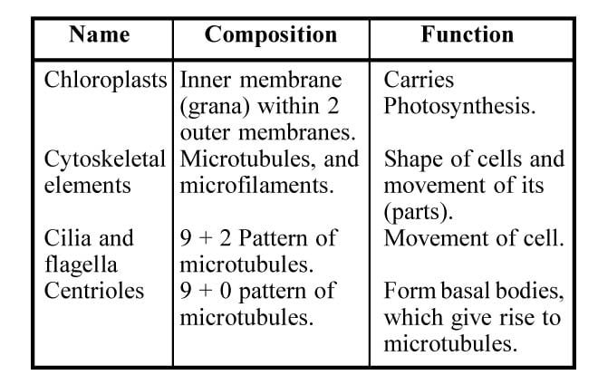
Chloroplast : Chloroplasts are the green coloured plastids containing chlorophyll pigments. Chloroplasts are found in green parts of plants such as leaf, young stem, calyx of a flower, young fruit, etc. The green pigment chlorophyll is responsible for trapping solar energy and converting it into chemical energy (food material) in a process known as photosynthesis.
Chromoplast : Chromoplasts are plastids which have many colours but not green. These plastids are found in the cells of flower-petals, fruit skins, coloured leaves, etc. Bright colours of petals of flowers are due to chloroplasts. These petals attract insects which carry-out pollination.
Leucoplast : Leucoplasts are also plastids but they are without any colour and pigment. Leucoplasts are found in underground roots and stems which do not receive exposure to sunlight. Leucoplasts of the cells of the roots and underground stems change the sugar into starch. Leucoplasts are involved in the storage of food materials in the cell.
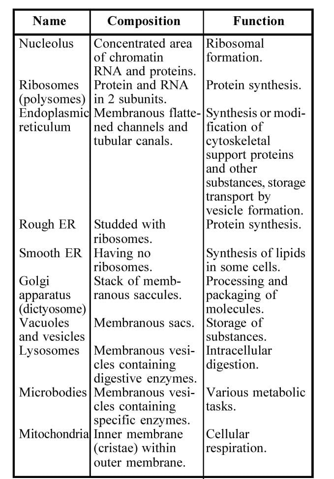
Mitochondria : Mitochondria are cell organelles associated with the release of energy from food materials (respiration) are knwon as ‘mitochondria’ (singular—mitochondrion). Mitochondria are found in all plant and animal cells. These are not found in bacteria and blue-green algae. The mitochondria may be considered as the power store house of the cell, because they are the aerobic respiratory centres of the cell. The mitochondria mainly are energy converters.
Golgi body or golgi apparatus : The golgi body or golgi apparatus are the cell organelle occurring near the nucleus and associated with the secretory activity of a cell. Golgi body is present in all plant and animal cells and it appears near the nucleus. The main function of golgi body is not yet fully understood. The secretory activity of the cell is carried out by golgi body. Metabolism of fats is also carried out by golgi bodies. It also plays a role in the metabolism of fats. It is also associated in plant cells with cell plate formation.
Lysosomes : The lysosomes are tiny, submicroscopic sac-like structure containing digestive enzymes. Lysosomes have been found in many animal cells and in the meristematic cells of a few plants. They are involved in many functions including a role in digestion of food particles, in defence against bacteria and viruses, in destroying worn out organelles of the cells often resulting in death of the cell. Due to this last role, they are referred to as suicide bags of the cell.
Endoplasmic reticulum : Endoplasmic reticulum is a system of folded membrane in the cytoplasm which functions as site of protein synthesis and transport. It is found everywhere in all eukaryotic plant and animal cells. They are distributed near the nuclear membrane. They connect nuclear membrane with the plasma membrane. They are distributed widely throughout the cytoplasm. The Endoplasmic reticulum gives a large surface area upon which numerous biochemical reactions can occur.
Ribosomes : These are extremely minute granular, and spherical structures present in the cell. Ribosomes are present in plant and animal cells everywhere. Ribosomes are the main places of protein synthesis in the cell.
The Cytoskeleton : The cytoplasm of eukaryotic cells has an extensive network of minute tubules and filaments, called microtubules and microfilaments. To maintain the shape of the cell, microtubles and microfilaments form the structural frame work within the cell. These are known as the cytoskeleton.
Microfilaments : These are very fine protein filaments about 7 nm in diameter. They are present in all eukaryotic cells and consist of the protein actin. They are perhaps involved in endocytosis and exocytosis.
Microtubules : Microtubules are present in all eukaryotic cells and are hollow unbranched cylindrical organelles. They are very fine tubes. They are involved in formation of centrioles, cilia and flagella.
Centrioles and centrosome : These are the non-membranous small hollow cylinders (about 0.3-0.7 mm long and about 0.2 mm in diameter) that occur in pairs in a distinct region of the cytoplasm. The centrioles are found in pairs at right angles to one another near one pole of the nucleus. The cytoplasmic matrix that holds the centrioles is called as centrosphere. The paired centriole is known as diplosome. These are found in most animal and lower plant cells. The centrioles form spindle fibres. In cell division they help in the movement of chromosomes.
Cilia and flagella : Cillia are minute hair-like protoplasmic processes extending from the cell surface. Microscopic whip-like protoplasmic extensions of the cell surface is termed ‘flagellum’ (plural-flagella). These specialized surface structures present in eukaryotic cells and also in prokaryotic cell like bacteria.
Vacuole : Vacuoles are known as the membrane-bound non-living cavities in the cytoplasm that contain watery cell sap. These are found in both plant and animal cells. The vacuoles act as storehouse of various secretory, excretory and food materials. In the cell, they provide space for water storage.
Ergastic substances : These are the non-living metabolic waste products of a cell which remain dissolved in cell sap. They also remain distributed in the cytoplasm as solid particles. These substances play an important role in the physiological processes of plant. These ergastic substances are used by man as economic products.
Cellular Respiration
Cellular respiration : Cellular respiration is a biochemical as well as an energy releasing process. Commonly it takes place within the living cell. In this respiration oxygen is absorbed and carbondioxide is released. Respiratory substrates (food are oxidised with the help of this oxygen and carbon dioxide is produced).
Types of Respiration
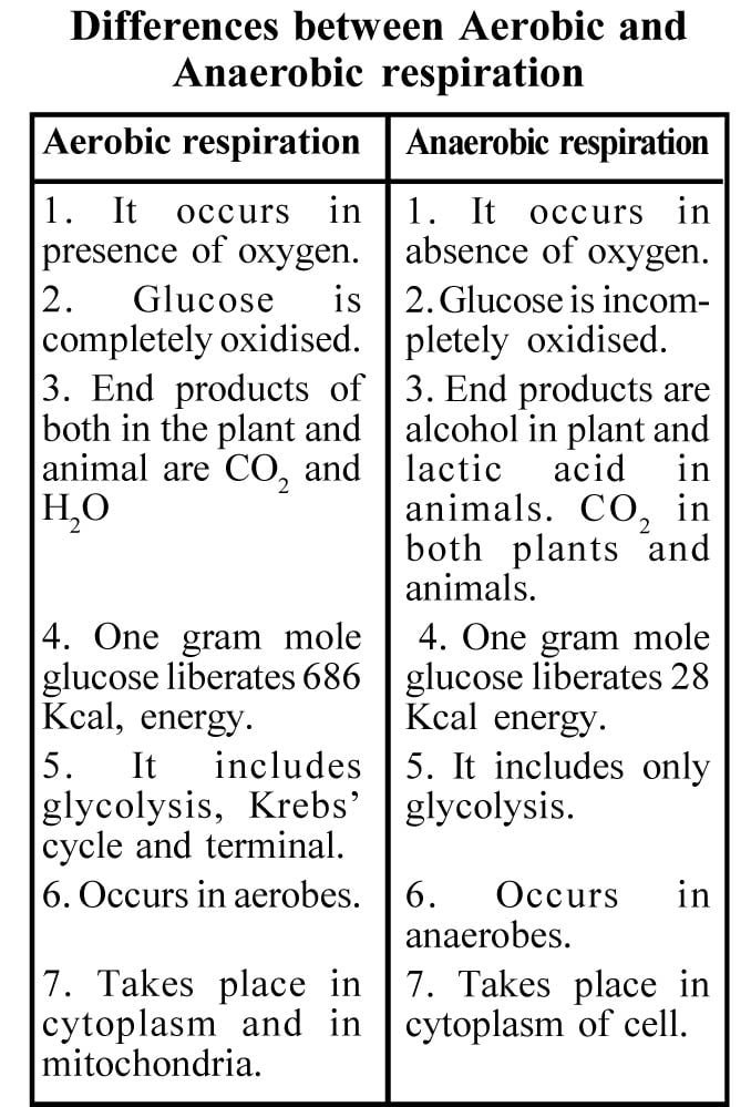
Respiration is mainly of two types. These are known as aerobic respiration and anaerobic respiration.
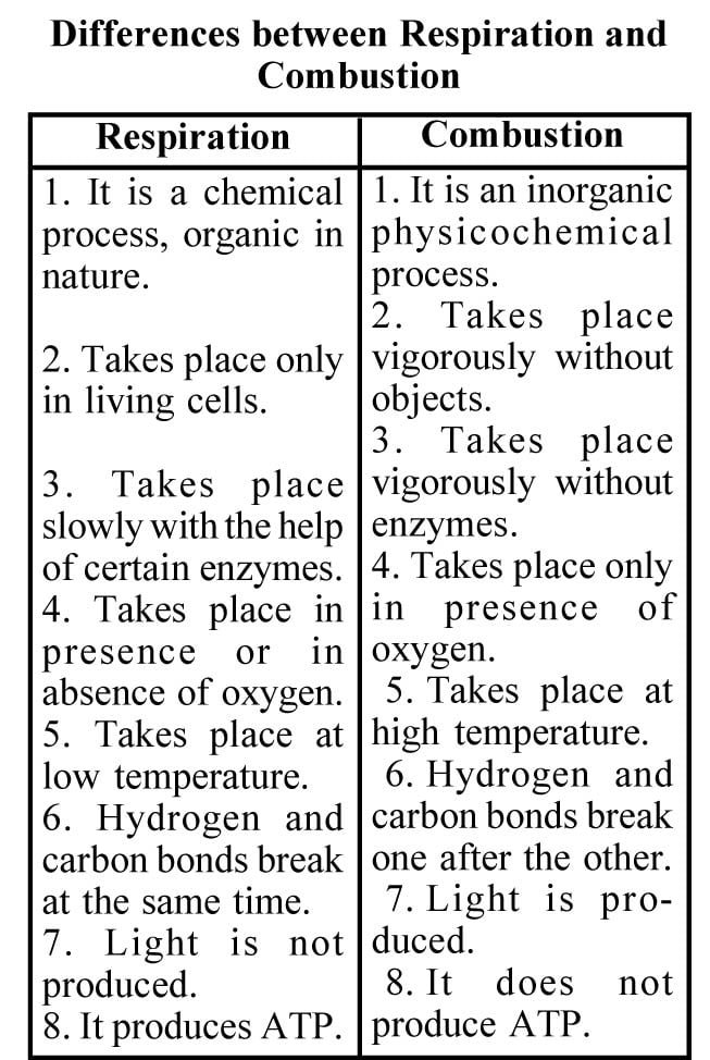
Site of aerobic respiration : Aerobic respiration takes place in the living cells at places which need oxygen. These are called aerobes.
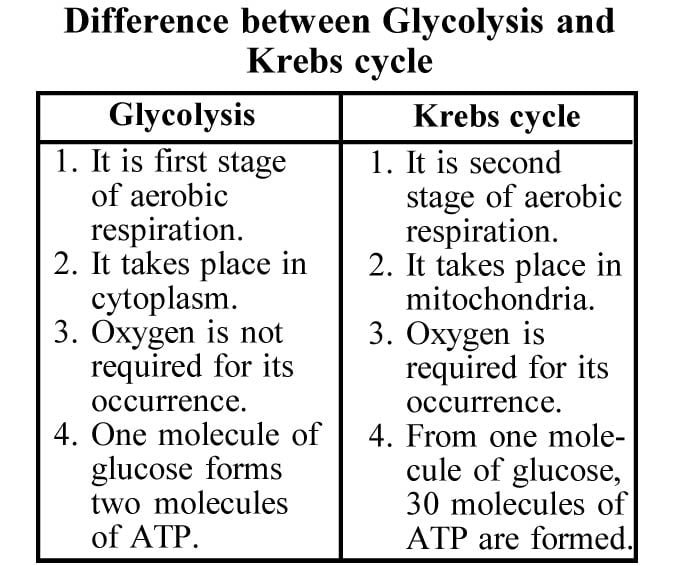
Mechanism of aerobic respiration : Glycolysis and krebs cycles are two stages by which the aerobic respiration is completed in the living cells. Glycolysis is called first stage and krebs cycle is second stage.
Site of anaerobic respiration : This type of respiration generally takes place in the anaerobic bacteria (e.g., Clostridium, Lactobacillus), unicellular fungus (yeast), and in plant seeds, parasitic animals (tape worm, round worm, Monocystis, etc.), in the skeletal muscle fibres (cells) during vigorous exercise and in the cytoplasm of the general cell.
Mechanism of anaerobic respiration : Anaerobes means organisms which can live without oxygen. This type of respiration can take place in the organism which can live without oxygen.
In this type of respiration only the process of glycolysis takes place. Krebs cycle does not take place because there is no oxygen.
Economic importance of fermentation : Fermentation is a very important process for making alcohol, beer, vinegar, curd, lactic acid etc. So it is useful in industries which manufacture these products.
Cell Division
Cell division is responsible for the growth and reproduction of the organisms.This is a continuous process which takes life long. Rudolph Virchow (1859) first proposed that new cells are formed from pre-existing cells by division. This is known as cell lineage theory.
Cell divisions are of three kinds, namely—(1) Amitosis (2) Mitosis and (3) Meiosis.
1. Amitosis : This type of cell division occurs in bacteria, yeast, Amoeba and tapetal cells. It does not show the formation of spindle apparatus and is primitive division.
In Amoeba, a well defined true nucleus can be seen. During the division, the nucleus on elongation changes to dumbbell shaped. It finally splits into two daughter nuclei. This is followed by the formation of a constriction which becomes deep and ultimately splits the cell into two daughter cells. This results in increasing the number of cells.
2. Mitosis : This type of cell division occurs in the vegetative cells. This is also called as ‘somatic division’. In somatic division the mother cell produces two daughter cells which are identical in shape, size and character. Therefore, it is called ‘equational division or homeotypic division’. Process of mitosis causes increase in cell number and increases the growth of the organism.
3. Meiosis : This kind of cell division occurs in the reproductive cells or germinal cells. In meiosis the mother cell produces four daugher cells called ‘gametes’ in which the number of chromosomes is reduced to half. Hence it is called ‘reduction division.’ The gametes carry genetic recombinations and therefore are unidentical with that of the mother cell. That is why this division is called ‘heteroptypic division.’ This process is highly helpful in the reproduction of organisms.
Mitosis or Equational Division
This type of division occurs in the somatic cells. It can be observed in apical meristems of root and stem. This division makes the size increase, change shape and volume of plant parts and causes growth.
Stages of Cell Cycle
Cell cycle has two stages
(A) Interphase or Resting Phase
(B) Mitotic phase.
(A) Interphase : There are three distinct stages of inter-phase called G1 phase, S-phase and G2 phase. This subdivision has been done on the basis of biochemical studies. These three phases stand-G for gap period and S-for synthesis.
(1) G1 phase : During G1 phase the cell increases in size. A lot of RNA and protein is synthesised in this stage.
(2) S-phase : During S-phase, the DNA present in each chromosome is duplicated. This is followed by the splitting of chromosome arm into two chromatids which are united at centromere.
3. G2 phase : During G2 phase the synthesis of protein and RNA is continued.
(B) Mitotic phase : At the end of interphase, the cell gets ready for division. In mitotic phase the nucleus divides first and it is followed by division of cytoplasm. Nuclear division is called ‘karyokinesis’ and cytoplasmic division is called ‘cytokinesis’. These two are given below :
(a) Karyokinesis : The dividing nucleus involves a number of sequential changes. Karyokinesis is further subdivided into four phases called Prophase, Metaphase, Anaphase and Telophase.
(1) Prophase : Prophase is the first stage of nuclear division. In the beginaing of this phase, the prochromosomes are long, slender and they spread extensively in the nucleoplasm. They gradually become short, thick, stout and develop into rod shaped chromosomes by coiling process. The chromosome arm appears longitudinally and split into two parts called chromatids which are united at the centromere.
In the last prophase, the nuclear membrane dissolves and the nucleolus decreases in size and finally disappears. The chromosomes are randomly scattered in the cytoplasm.
(2) Metaphase : In metaphase fusion of microtubules forms bipolar apparatus. They attach themselves to the kinetochore part of the chromosomes and bring them to the equator of the cell where they are oriented in a systematic manner. Thus, all the centromeres lie in the same plane forming the equatorial plate. The chromosome arms hang freely in the cytoplasm.
(3) Anaphase : In anaphase, the spindle fibres begin to contract causing pressure on the centromeres. As a result the centromere of each chromosome divides and thus, the two chromatids are separated. The chromatids with their own centromeres are now called as ‘daughter chromosomes’. The spindle fibres pull the daughter chromosomes to the poles. During the movement of the chromosomes to the poles, the centromeres lie ahead followed by arms. The chromosomes appear in the shape of V, L, J or I.
(4) Telophase : In telophase the daughter chromosomes arrived at the poles become thin, long and lose their visibility due to despiralisation to form chromatin. The nuclear membrane reappears and the nucleoli are organised again from their relevant chromosomes. At the end of telophase, two independent daughter nuclei are organised. The daughter nuclei then enter the G state of cell cycle.
(b) Cytokinesis : This occurs after telophase. At the end of the telophase when the daughter nuclei are formed the spindle fragments gather at the equator region and form a barrel shaped structure called ‘phragmoplast’. The vacuoles of golgi apparatus enter the phragmoplast and release pectins into it. Thus a liquid form of cell plate is formed. The cell plate grows and gets connected with the parent wall. It gradually undergoes physical and chemical changes to form middle lamella. This divides the cytoplasm into two parts. On both sides of the middle lamellum the primary wall materials like cellulose and semi-cellulose gets deposited. Finally two daughter cells are formed.
Significance of Mitosis :
1. Mitosis causes growth in the organism.
2. The daughter cells formed by mitosis resemble the mother cell in qualitative and quantitative characters. Hence, it is important in conserving the genetic integrity of the organism.
3. Mitosis is responsible in unicellular organisms for reproduction.
4. It helps in the rear and tear mechanism of the plant body and also useful in the healing of wounds.
5. Mitosis is highly useful in the regeneration of lost parts and for grafting in vegetative reproduction.
Stages of Meiotic Cell Division
Meiotic cell division occurs in two stages called (1) Meiosis-I (2) Meiosis-II.
Meiosis-I is a heterotypic division. In this division, the diploid parent nucleus divides into two daughter nuclei each having haploid set of chromosomes. Meiosis-II is homeotypic division. In this the two haploid nuclei divide mitotically and form four haploid cells. Thus, from a single parent cell containing diploid (2n) chromosomes, four haploid (n) daughter cells are formed.
Meiosis-I has four phases known as :
    (A) Leptotene
Prophase-I ® Again it (B) Zygotene
has five
substages
Metaphase-I (C) Pachytene
Anaphase-I (D) Diplotene
Telophase-I (E) Diakinesis Meiosis-II also has four phase known as :
Prophase-II 2. Metaphase-II
Anaphase-II 4. Telophase-II
Meiosis-I (or) First Meiotic Division : In the first meiotic division the nuclear division takes place in four main stages—1. Prophase-I
Metaphase-I 3. Anaphase-I and
Telophase-I.
Prophase-I : For a systematic study, prophase is divided into five sub-stages discussed as (A), (B), (C), (D) and (E).
(A) Leptotene : In this phase the nucleus becomes bigger in size. The chromosomes are long and slender and show bead like structures. The number, size and position of chromomeres is constant and helps in identifying the chromosomes. The chromomeres are considered to be active genetic centres. The chromosomes are arranged parallel and well-separated but at the end of Leptotene, the homologous paternal and maternal chromosomes come close together at a point. This condition is known as ‘bouquet stage’.
(B) Zygotene : In this, the similar paternal and maternal chromosomes attract each other and make pairs. These are called ‘bivalents.’ The pairing process is known as ‘synapsis’.
Synapsis is completed by three methods viz.
(i) Proterminal : Pairing starts at the polarised ends and goes gradually towards other extremity.
(ii) Procentric : Pairing starts at the centromere and goes towards the end of the chromosomes.
(iii) Random or intermediate : Pairing occurs simultaneously at various places along the length of the chromosomes. The process is random.
In Zygotene enlargement of nucleous and the initiation of spindle formation takes place.
(C) Pachytene : In each chromosome pachytene is characterised by the appearance of two chromatids. Thus, in each bivalent, we can see four chromatids. These are called ‘pachytene tetrads’. The chromatids of each similar chromosomes are called ‘sister chromatids’ and those belonging to other chromosomes are called ‘non-sister chromatids’. The exchange of non-sister chromatids takes place mutually at one, two or many places. Such points where the chromatids physically contact with each other are called ‘chiasmata’. At this stage the chromosomes appear in ‘X’ shaped structure. During the formation of chaismata, the chromatid arms first break due to the action of an enzyme called ‘endonuclease’. The broken chromatid arms mutually exchange with each other and get united by the action of ‘ligase’ enzyme. The formation of chiasmata leads to the exchange of genetic material and results in the recombination of characters. This is known as ‘crossing over’. It gives rise to the origin of new species and thus leads to evolution.
(D) Diplotene : In diplotene similar chromosomes repel with each other due to the weakening of synaptic forces. In diplotene mainly the condensation, contraction and thickening of the chromatids take place.
(E) Diakinesis : The chiasmata starts moving towards the chromosome ends. This displacement is known as ‘terminalisation’. The bivalents become very thick and short and move to the periphery of the nucleus. The nucleolus starts to disappear, the nuclear membrane disrupts and the chromosomes are released into the cytoplasm. The chromosomes remain far apart from each other as much as it is possible.
Metaphase-I : In metaphase-I, bipolar spindle apparatus appears. The chromosomes move to the equator of the cell and arrange themselves with their centromeres directed towards the opposite poles and their arms towards the equator. The homologous chromosomes get fused by the chiasmata at the telomeric ends.
Anaphase-I : Anaphase-I is characterised by the movement of daughter chromatids of each homologue united by their centromeres towards their respective poles with the help of spindle fibres. This is known as ‘segregation of chromosomes’. Thus the two sets of chromosomes are separated and carried to the opposite poles.
Telophase-I : In telophase-I the chromosomes arrived at the poles begin elongating by lessening their coils. The nuclear membrane and nucleolus reappear. Thus two daughter nuclei are formed, each possessing a haploid (n) set of chromosomes.
Meiosis-II (or Second Meiotic Division) : It resembles the mitotic division. Each daughter nuclei divides to form two cells without any change in the number of chromosome numbers. It has four stages called prophase-II, metaphase-II, anaphase-II and telophase-II.
Prophase-II : In this phase nuclear membrane and nucleolus disappear and chromosomes are organised. Each chromosome exhibits two chromatids.
Metaphase-II : In Metaphase-II two spindle apparatic are made perpendicular to the one formed at the metaphase-I. On the equatorial plate the chromosomes are arranged.
Anaphase-II : The centromeres are divided and form daughter chromosomes. The spindle fibres pull the chromosomes towards the opposite poles.
Telophase-II : The daughter chromosomes arrived at the poles become thin and transform into chromatin. Nucleolus and nuclear membrane are reorganised. We see that at the end of telophase-II, four daughter nuclei are formed.
Cytokinesis : Cytoplasm division can occur after each nuclear division. It can be delayed until four daughter nuclei are formed. The process of cytokinesis is the same as that of mitotic division.
Thus in meiosis each mother cell splits into four daughter cells.
Significance of Meiosis :|
The process of Meiosis helps in the maintenance of a constant number of chromosomes from one generation to the next.
Due to crossing over, genetic recombinations is caused which help in the origin of new species. It lead to evolution.
It helps in the formation of the gametes. It is very useful in sexual reproduction.


