The cell is the basic unit of the structure of all living beings.
The study concerning cell is known as cell biology.
All living beings-animals and plants are composed of cells.
The cells play a similar role in body building as the bricks play in the construction of a building or atoms play in the structure of matter.
History
Van Leeuwenhock (1632) is known as the father of cell biology because he could see protozoa, bacteria etc. with his primitive microscope. Robert Hooke modified the microscope in 1665 and studied cells. Cell biology became an important field in the beginning of 19th century.
Details about the cells are given below—
Cell : It is a fundamental structural and functional unit of all living organisms.
Cytology : Cytology is the branch of biology pertaining to the study of the cells as viewed through a microscope. Now-a-days the term cell biology is used.
Cell Biology : It is the branch of biology dealing with the study of cell and its components by the techniques borrowed from different branches of sciences.
The Cell Theory : The 19th century was an era in which the cell was investigated extensively with the microscope. In 1829, a Frenchman, H.J. Dutrochet boiled some tissues in an acid and separated the cells. German Scientists, M.J. Schleiden (1938) and T. Scllwann (1839) were credited for proposing the cell theory that all the animals and plants are composed of cells. Another German scientist, Rudolf Virchow, provided strong evidence for the idea that every cell is derived from a pre-existing cell by some types of division.
Putting all these observations together, the modern cell theory can be stated as follows :
(i) Origin, nature and continuity of life bound up in the cell.
(ii) All organisms are composed of cells or cell products.
Cell Portrait : The cell is the ‘biological unit’ of activity with a small mass of protoplasm. They have the machinery for day-to-day maintenance of a living body and for the production of more units like themselves. Therefore, a cell in a more proper way may be defined as “the smaller organized unit of living matter (protoplasms), which is capable of prolonged independent existence and replacement of its own substance (growth and self reproduction) in a suitable environment. The unicellular organism is represented by a single cell, whereas many cells enter into the formation of a multicellular organisms.”
Modern definition of a Cell : It is defined as a piece of nucleated cytoplasm surrounded by a cell-wall or membrane, existing singly or in groups and containing structures of various kinds. Cells are of two types —
Prokaryotic Cells : These cells do not possess well-formed nucleus enclosed in a nuclear membrane. These cells, instead of a nucleus, have a nuclear zone called nucleoid. These cells either lack all other organelles or have them in a primitive undifferentiated condition. The functions of respiration and phtosynthesis are carried out by the cell membrane and its infolding. Cells of blue-green algae, bacteria and virus are of this type.
Eukaryotic Cells : These cells have definite nuclear membranes forming two distinct compartments of the cytoplasm and the nucleus. In these cells, respiration, excretion, photosynthesis and metabolism take place in definite cytoplasmic organelles. This type of the cells are found in all the higher animals and plants, protozoa, fungi and majority of algae.
Morphology of Cell
Size : The size of different cells ranges within broad limits. Some plant and animal cells are visible to the naked eye, such as egg of certain birds have a diameter of several centimeters (Egg of ostrich is as big as 170 × 155 mm). But majority of cells are visible only under microscope.
The smallest living cell is that of bacterium of pleuropneumonia, called PPLO (Pleuropneumonia, like organi-sms) which measures 0.1 to .05 micron in size. Organisms like Pleuropneumonia are also called elementary bodies. The long nerve is also an example of the extremes in cell sizes, the range of which is quite remarkable in the living world.
In the human body, nerve cells are often about 90 cm in length. In plants certain algae have gigantic cells. An algae Acetabularia consists of a single cell about ten centimeter in length. The size, specially the surface, plays an important role in relation to the function of the cell. A cell takes nutrients from its environment and gives off wastes through its surface. The factors governing the size of the cell are : (i) the nucleocytoplasm ratio, or the ratio between the volume of the nucleus and the cytoplasm, (ii) the ratio of the cell surface to the cell volume, and (iii) the rate of metabolism. It is now known that the size of cell does not show any relation to that of the body size of an organism as the elephant, whale and camel do not have large cells.
Shape : Cell has numerous shapes. The cells of certain unicellular organisms, such as Amoeba, Diatoms, Acetabularia and Bacteria exhibit a number of shapes.
Among the cells of multicellular forms, a variety of shapes are present in the same organism. The shape of a cell depends mainly on functional adaptations and partly on the surface tension and viscosity of the protoplasm, the mechanical action exerted by the adjoining cells and rigidity of the cell membrane.
The body of unicellular organisms consists of a single cell. Most of the animals and plants have many cells in the body and are known as multicellular animals or plants. The cortex of the human brain may consists of nine billion two hundred million cells. The body of an average adult man is made up of about 100 trillion (10,00,00,00,00,000,000) cells.
Physical Structure
The animal cells are different from plant cells. In spite of these differences, all of them may show some common features. A typical cell consists of two main components—the cell membrane and protoplasm; the latter is further differentiated into the nucleus and the cytoplasm. Cell of micro-organism like bacteria, viruses and blue green algae do not possess definite nuclear membrane and membrane organelles, and it is therefore known as ‘prokaryotic cell’.
The cells of other organisms contain definite nuclear membrane and are thus called enkaryotic cells. Cell membrane is the outer covering of cell enclosing the internal parts and allow some materials to pass in and out of the cell; acting as selective membrane. The cell membrane in animal cell contains lipid and protein and is thus called plasma membrane while in plants it is known as cell wall containing cellulose. In cytoplasm certain small membrane-bounded bodies which are called organelles are lying embedded. These bodies or organelles are the mitochondria, golgi bodies, endoplasmic reticulum, lysosomes, ribosomes, plastids etc. Briefly, mitochondria is the power house of cell and controls oxidative activities, plastids in plant cells perform several functions like photosynthesis, storage of reserve food or colour in plant materials; Golgi bodies secrete enzymes and hormones; Lysosomes are capable of digesting extra as well as intracellular particles.
Enkaryotic cells contain definite nucleus which contains thread like bodies, the chromosomes. Each chromosome is biochemically made up of a chromatic material. Chromosomes bear genes which transmit parental characters to the offspring. Gene contains the hereditary material the DNA (deoxyribo-nucleic acid).
Chemical Nature of Protoplasm : The chemical components of the cell can be classified as inorganic (mineral salts) and organic (proteins, carbohydrates, nucleic acids etc.) substances. Although the most prominent constitutent of protoplasm is water which gives to the protoplasm its characteristic feature. Lipids are important as all membranes and carbohydrates serve as nutrient stores. The following table gives approximate figures of the chemical components present in protoplasm :
Substance Percent
1. Water 85%
2. Protein 10%
3. Lipid 1-3%
4. Carbohydrates 1-2%
5. DNA 0.4%
6. RNA 0.7%
7. Other organic materials 0.4%
8. Inorganic substances 1.5%
Cell Wall and Plasma Membrane : The plant cells contain a definite rigid envelope called the cell wall. The cell wall is the outer rigid and protective layer around the plasma membrane which provides the mechanical support to the cell. The cell wall also determines the shape of plant cells. Cell wall plays a major role in helping the aerial portion of the land plants to withstand gravitational forces.
Structure of the cell wall
Cell Wall : The plant cells are surrounded by a rigid non-living and thick covering above their plasma membrane called cell wall. There are three cell walls—
(i) Primary wall : The outer most layer is called the Primary Wall. It is formed first. It is thinner than the secondary wall and in it the fibrils are found running in many direction. The primary wall of the adjacent cells is joined with each other with the help of meddle lamella.
(ii) Secondary wall : Below primary layer lies the secondary wall forming layer. In each layer, the individual cellulose fibrils are oriented parallel to one another.
(iii) Tertiary wall : The inner most layer of cell wall is called the tertiary layer. It may or may not be present, if present it contains an additional chemical substance: the xylem.
Chemical nature of cell wall : The cell walls of plant cells are composed of carbohydrates known as cellulose which contains long chains of glucose molecules (3000 molecules). Many chains of cellulose molecules lie parallel to each other to form the elementary fibrils known as micelle. Twenty such micelle when get arranged parallelly, form micro fibrils.
These micro fibrils form large sized bundles of cellulose fibres to form the macrofibrils. The macrofibrils consist of many cellulose fibrils and form the main frame work of the cell wall. The fibrils contain additional compounds such as lignin, pectin and cutin etc.
Plasma Membrane
It is also known as cell membrane and is defined as the outer boundary of the cytoplasm of the cell which forms an important barrier between the cytoplasm and the outer environment of the cell. It is a living and dynamic membrane.
Structure of the Plasma Membrane : The plasma membranes of the cell are composed of proteins and lipoids. Danielli and Hugh Davson proposed that that plasma membrane consisted of three layers; a middle double molecular layer of phospholipids and two protein layers on either side of it. Each phospholipids had two ends, one phydrophobic and other hydrophilic. The total thickness of plasma membrane is 75 Å. Each protein layer is about 20 Å in thickness and the phosopholipid is 35 Å thick.
Functions of Plasma Membrane
The function of plasma membrane is to regulate the flow of meterials into and out of the cell. This can be done by the following methods.
(i) Passive transport : In passive transport there is a movement of molecules or ions from higher concentration to lower concentration. It is similar to that of osmosis. The entry of water into a cell is termed as endosmosis; water exit is called exosmosis.
Active Transport : In active transport there is a movement of molecules or ions from lower concentration to higher concentration with the utilization of energy. It has been suggested that a carrier molecule exists in the membrane itself. This picks up Sodium from outside forming a temporary complex and carry it inside the cell, where it is released. On its way back, it may transport potassium ions outside the cell. During the movement of sodium across and potassium the membrane energy is required.
Pinocytosis and Phagocytosis : In some cases certain particles can neither get in nor come out of the plasma membrane. In such cases plasma membrane adopts special methods like endocytosis where the substances are taken in by the plasma membrane and exocytosis where the substances are thrown out by the cell. In endocytosis if the solid foreign particle bodies are taken in and engulfed the phenomenon is called the phagocytosis and the term pinocytosis is applied where fluid substance in large amount is taken in by the plasma membrane.
During phagocytosis pinocytosis and vacuoles are developed which either contain solid particles or fluid substances and are called phagosomes or pinosomes, respectively.
Endoplasmic Reticulum and Ribosomes : The endoplasmic reticulum is present in all the cells but very often absent in egg and in embryonic or undifferentiated cells but increases with differentiation. The spermetocytes have poorly developed endoplasmic reticulum.
The adipose tissues engaged in lipid metabolism contain only smooth endoplasmic reticulum.
The cells of those organs which are actively engaged in the synthesis of protein, as the pancreas and other endocrine glands, are found to contain highly developed rought endoplasmic reticulum.
In the striated muscle, the endoplasmic micreticulum takes a special form and is known as sacroplasmic reticulum. The liver cells possess both types of endoplasmic reticulum.
Ribosomes : They are spherical bodies which are about 150-200 Å in diameter. Each ribosome consists of two distinct subunits of unequal sizes. Ribosome functions as the site of protein synthesis.
In cells a good number of ribosome is present. In all rapidly growing cells of animals, plants and bacteria a large amount of ribosome is present. In growing E-coli about 20,000 to 30,000 ribosomes have been counted. However, if the rate of protein synthesis is slowed down, the number of ribosome is dropped automatically.
Structure : In the electron micrographs, ribosomes are seen as spherical bodies, roughly about 150 Å to 250 Å in diameter.
Each ribosome consists of two distinct sub-units of unequal sizes forming cap and dome shaped structures. These sub units are further composed of protein and R N A. in the ratio of 40 : 60 respectively.
Class of ribosomes
Three classes of ribosomes have been studied so far.
(i) Animal cytoplasmic ribosomes.
(ii) Plant cytoplasmic ribosomes.
(iii) Organelle and bacterial ribosomes.
Ribosomes play an important role in protein synthesis. During protein synthesis some of them give the appearance of beads along with m RNA thus forming polyribosomes.
Golgi Apparatus : It is an organelle which consists of a highly organised system of membrane plates and tubules. The main function of Golgi appratus is the secretion of cell wall material in plants. The size is large in the nerve and gland cells and small in muscle cells. In general, the Golgi apparatus is well-developed while the cell is in active state. When the cell grows old, the apparatus progressively diminishes in size and disappears.
Detailed Structure : The structure of Golgi apparatus under the electron microscope appears as given below :
1. Flattened sacs (cisterne)
2. Large vacuoles
3. Clusters of small vesicles.
The flattened sacs are quite similar in appearance to the smooth shaped endoplasmic reticulum. It is seen that during the period of rest the Golgi bodies are flattened. On being filled up with secretion they assume spherical shape. The small vesicles are pinched off from the end of sacs.
Morphology : Golgi body occurs in almost all enkangotic cells. Their number per plant cell can vary from several hundred as tissues of corn root.
1. Role in Secretion : Golgi apparatus is considered to play an important role in the secretory functions of a cell. Enzymes and hormones are the secretory products of Golgi apparatus. Besides this, it also has some role in the synthesis and secretion of polysaccharides, phospholipids and pigment formation.
2. Role in Vitellogenesis : In the early oocyte, the Golgi apparatus forms yolk nucleus along with mitochondria and endoplasmic reticulum. The yolk nucleus possesses rings of the organelles listed above and thereafter produces yolk droplets.
3. Role in Spermatogenesis : Golgi apparatus develops acrosome during spermatogenesis. An acrosome is a structure located at the tip of sperm which is used in the penetration of sperm during fertilization.
4. It helps in the formation of cell wall in plant cell during division.
Microbodies : Microbodies are minute bodies bounded by single membrane having specific roles and unique characteristics. These organelles have a central granular or crystalliod core containing some enzymes. The well known microbodies are the lysosomes, peroxisomes and spherosomes.
Lysosomes : It is a membrane-enclosed vesicle containing digestive enzymes. It functions in intracellular digestion.
Lysosomes are bag like small bodies; surrounded by a membrane. They are comparatively small organelles, on an average, 0.5 micron diameter.
They contain nearly 40 different acid hydrolyses, one of the most important enzymes being the acid phosphatase.
Usually the lysosomes occur in most animal cells and in a few plant cells. They are particularly numerous and large in cells such as macrophages which perform special digestive functions.
Peroxisomes : These are small organelles which are bound by a single unit membrane. These have a central core called nucleoid and are closely associated with endoplasmic reticulum in the cell. These are involved in the photorespiration of the plant cells and lipid metabolism of the animal cells.
Spherosomes : These are small spherical particles which have high deposits of fats and lipids. These originate from endoplasmic reticulum. These contain some enzymes like acid phosphatases which do not show much to the lytic activity.
Glyoxysomes : These are the microbodies in plant cells which are involved in the glyoxylate cycle. When germination of seeds takes place, glyoxylate cycle operates by which stored fat is converted into carbohydrates.
Mitochondria : Mitochondria is composed of layered membranes enclosed by a separate outer membrane, it is the chief site of respiratory (energy releasing) metabolism and ATP (adenosine triphosphate) formation. Mitochondria is of universal occurrence, being present in both plant and animal cells with exception to bacteria and red blood corpuscles. The number of size of mitochondria depends upon functional state of cell. The size of mitochondria also varies from cell to cell again depending upon the functional state of cell. They generally attain 5 to 10 microns length and 0.5 to 1.0 micron width. The largest mitochondrion is found to have a length of 40 microns. The oocytes of amplubia contain 20 to 40 microns long mitochondria.
Structure : Electron microscopic studies have clearly shown that the mitochondrion possesses a double membrane: an outer limiting membrane and an inner limiting membrane. Each membrane is about 60 Å and remains apart from each other by a space of 60 Å to 100 Å width. The space is called intermembranous space. The outer membrane is continuous and uninterrupted while the inner one shows extensive foldings. The foldings are the cristae which increase the total surface areas of the inner membrane. The space bounded by the inner membrane forms inner compartment and remains filled with a dense material known as mitochondrical matrix. The space within the cristae which is continuous with the permitochondrial space is known as intracristae space.
The inner surface of inner membrane and the outer surface of outer membrane are deposited with thousands of evenly space minute particles of 90-100 Å diameter. These particles are called the mitochondrial particles.
Mitochondria contains about 25 to 35% lipid, 5 to 7% RNA, 60 to 70% protein, circular DNA and nearly 60 different enzymes.
Functions
1. It helps in cellular respiration.
2. It forms mitochondiral spiral in the collar region of sperm.
3. During oogenesis it synthesizes yolk particles.
4. As the name power house indicates, it stores and liberates energy (ATP) during metabolic activities in the cell.
Chloroplasts : These are the most important cytoplasmic organelles of the cells of all green plants and some protozoans such as Euglena. They convert the solar energy into chemical energy. A chloroplast is a form of plastid. There are several kinds of plastids which are classified into three groups. The leucoplasts or the colourless store reserve food material; the chloroplasts give green colour to plants and, the chromoplasts contain colour other than green such as brown, red, yellow etc.
Grana : The internal membranes of chloroplasts of eukaryotic cells are arranged in interconnecting stacks of membrane subunits. There stacks are called grana.
Stroma : The space between grana and membranes is called stroma.
Thylakoids : Each granum is composed of a stack of 10 to 30 disc-shaped membrane subunits called thylakoids. Each thylakoid is a thickened single membrane with the edges rounded off by proteins.
Photosynthesis : It is defined as the process which brings about the synthesis of carbohydrates like sugars from the two initial products, namely, carbon dioxide and water, in the presence of sunlight and chlorophyll. The summary equation of the photosynthesis is as follows.
6CO2+12H2O®C6H12O6+6H2O+6O2
Chemical analysis of chloroplast shows the following data :
Protein 50-60%
Lipids 25-35%
Chlorophyll 5-10%
Pigments like Carotenoids 1-2%
RNA = 2-3%
DNA =5%
Mg, Fe, Cu, Mn, = Traces.
Zn, and P
Functions : Chloroplasts carry out the most important function, the photosynthesis, and convert the kinetic solar energy into potential energy by synthesizing carbohydrates from CO2 and water.
Chloroplasts are presumed to participate in the synthesis of protein.
Chloroplasts contain different pigments and provide different colours to fruits and flowers.
Leucoplasts like proteinoplasts, chloroplasts and amytoplasts store plasts proteins, oils and starch respectively.
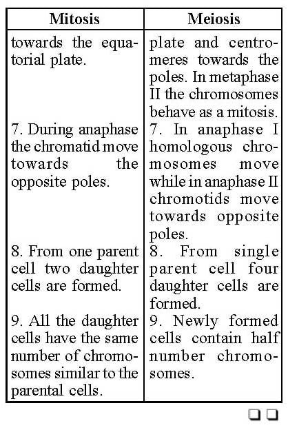
Centrioles and Basal Bodies : It is a structure composed of nine triplet sets of microtubules. It functions in spindle fibre organisation during cell division and as the basil attachment of cilia and flagella.
Basal Bodies : When centrioles produce flagella or cilia, they are known as basal bodies. Similar to centrioles, these have the same fundamental nine sets of triplet organisation.
Cilia and Flagella : These are the sepcial surface structures which are used as propellers in locomotion of the cells.
Detailed structure : Electron microscopic study of centriole shows that each centriole consists of nine sets of tubular structures of fibres and each of the latter is further made up of three or triplet strands or microtubules. These triplets are embedded in an amorphous matrix. In some cases all those triplets unite with each other by means of fine fibres. Some delicate fibres radiate from centre giving the appearance of cartwheel.
Biochemically centrioles may contain circular DNA.Functions
1. Forms cilia and flagella.
2. Helps in the formation of spindle during cell division.
3. Forms axial filament in sperm.
4. Helps in vitelloegenesis.
Cilia and Flagella : Cilia flagella are motile, hair-like appendages on the free surface of cells. These are cytoplasmic processes, which orginate from the basal bodies, embedded in the cytoplasm and extend out of it. In size these vary from 5 to 10 microns. Flagella in a cell can attain a size upto 150 microns. The diameters of both cilia and flagella are less then 0.5 microns.
HistoMorphologically cilia and flagella are similar. They resemble each other in having nine sets of tubules or peripheral fibres arrange in a circle.
Both cilia and flagella are chemically composed of 70 to 84% proteins, 13 to 23% lipids, 1 to 6% carbohydrates, and 0.2 to 0.4% nucleotides. The outer fibrils (doublets) contain a protein called tubulin which resembles closely with the action of the muscle cells. The arms and central fibrils contain the protein dynein which contains enzyme ATP, one of the cilia which split the phosphate bond of ATP to release energy.
Functions
1. They make organs of Locomotion of cell or organism.
2. The eggs of amphibians and mammals are driven out from the oviduct by the aid of vibratile cilia of the latter.
3. The cilia create food currents in lower aquatic animals.
4. In the respiratory tract, the ciliary movements help in the elimination of the solid particles from it.
5. Also perform sensory function.
Interphase Nucleus
Interphase Cell : It is the one when a cell is not in the process of cell division.
Nucleus : It is an organelle which is composed of a nuclear envelope enclosing the chromosomes and a nucleolus.
Nuclear Envelope : It is a structure of two membranes enclosing the nucleus; about 120-200 Å thick; contain pores about 200-700 Å in diameter.
Chromosome : It is a strand of genetic material located in the nucleus of eukaryotic cell. It is composed primarly of DNA and proteins. It contains genes in linear order.
Nucleolus : It is a dense nuclear structure which is formed by the aggregation of portion of chromosomes.
The most prominent particulate feature of a cell when viewed under the microscope is the nucleus.
In general, the nucleus tends to be spherical, but may be fusiform, flattened or ellipsoidal depending upon the cell shape and function. In leucocytes, cartilage cells and animal ova the nucleus becomes irregular or lobulated. The spinning glands of certain insect larvae the nucleus shows the complex branches. The size of the nucleus is variable from cell to cell. In general it bears some relation to that of the cytoplasm.
Nucleus is present in all eukaryotic cells. Generally the cells are uninucleate. But in some cases binucleate condition occurs. In a few others multinuclear condition exists (opalina). Nuclei are essential for the survival and long term continuation of the cells. If nucleus from any cell is removed, the latter becomes unable to survive for long.
The nuclear sap fills completely the nuclear space. The major chemical composition of the sap is protein and lipids.
Cell Division
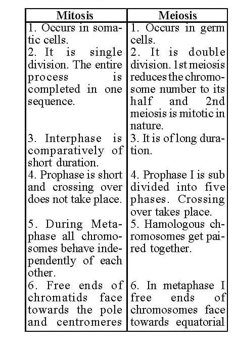
Mitosis : It is the usual process by which a nucleus divides into two. Four steps or phases of mitosis are as follows:
(a) Prophase : Initial stage of mitosis during which chromosomes appear within the nucleus.
(b) Metaphase : State of mitosis when chromosomes lie approximately at the right angle to the spindle fibre to which it is attached.
(c) Anaphase : Stage of mitosis when daughter chromosomes are separating towards poles of spindle.
(d) Telephase : Terminal stage of mitosis during which nuclei revert to resting stage.
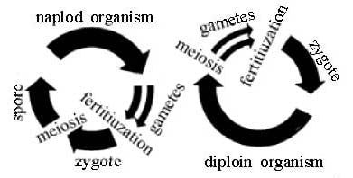
Meiosis : (Reproductive division) Two successive cell divisions of special kind starting in a diploid cell. Both divisions resemble mitosis but the chromosomes are duplicated only once before the first division, so that the number of chromosomes present in each of the four daughter cell is half that of diploid cell.
Genome : It refers to the basic set of chromosomes in the cells of an individual representing one copy of each chromosome. Thus a haploid cell has one genome whereas a deploid cell contains two genomes.
Polyteny : It refers to the basic set of chromosomes in the cells of an individual representing one copy of each chromosome. Thus a haploid cell will have one genome whereas a deploid cell contains two genomes.
Polyteny : It refers to the existence of many threads in a chromosome.
Spindle apparatus : It is the arrangement of microtubules radiating from two poles at opposite ends of the metaplate. Spindle apparatus consists of three types of fibres :
(a) Continuous fibres : These are not attached to the chromosomes but are extended from pole to pole.
(b) Chromosomal fibres : These are the fibres which are connecting the chromosomes to opposite poles of the spindle.
(c) Incomplete fibres : These are the fibres radiating from the pole. These are not attached to chromosomes and also do not reach the opposite pole.
A living cell is a partnership between the nucleus and cytoplasm and both are needed for the continued life and functioning of the cell and all the living organisms right from the fertilization of the egg or just after the development of shape and size. The growth of the organisms involves a multiplication of cells by divisions. In unicellular forms, this results in the multiplication of the organism themselves. In multicellular forms a new individual initiates a series of sub-divisions which will eventuate in many cells of the body or some.
New cells arise by the division of a pre-existing cell. Each daughter cell receives from its parent a complete set (genome) of hereditary information and sufficient organelles to be able to carry out its metabolic reactions. Each genome is packed into one or more chromosomes. Cells with single genome are called the haploid, those with two are called diploid; similarly those with more than two are known as polyploid.
Cell Division
Cell division can be conveniently described as:
1. Direct divisions
2. Indirect divisions
1. Direct divisions : It is also called mitosis division or fragmentation and is characteristics of the lower organism in which apparently no definite chromosomes are formed. In this the nucleus simply constricts and separates into two portions while in the metabolic condition no achromatic figure is formed. For example, in Amoeba or yeast or leucocytes of vertebrate the cell divides without the appearance of chromosomes.
2. Indirect divisions : Here the nucleus undergoes complicated changes before it is divided into daughter nuclei.
Indirect division involves both the division of the nucleus by a complicated process known as Karyokinesis and the division of the cytoplasm as the cytokinesis. In indirect division if the daugher cells contain the same number of chromosmes as their mother cells the division is thus called Mitosis division. On the other hand if the daughter cells contain half number of chromosomes the division is said to be the reduction division or Metosis.
Mitosis : Mitosis is the process by which one cell becomes two cells in so complicated but in a precise manner that each daughter cell possesses just the same amount of DNA and the same set of chromosomes that the mother cell possessed. It occurs in somatic cells. Cell division is a continuous and dynamic process but for the purpose of description it is generally divided into phases or stages.
(i) Interphase : The period of metabolic activity during which cell division is not in a process has been called the interphase or intermitotic phase. Interphase is the largest period in the cell cycle and may last for several days in cells. It is during this phase that the cell prepares to divide by duplicating its DNA and by synthesizing some micromolecules that will be required during mitosis. Interphase can be further divided into three sub phases (b) G1 phase (first growth or gap period).
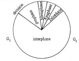
G1 phase includes the synthesis and organisation of the substrate and enzymes necessary for DNA synthesis. Therefore G1 is marked by the synthesis of RNA and protein (b) S phase (synthetic phase). In this phase the replication of DNA results in doubling the number of DNA strands in each chromosome. (c) G2 phase (second gap or second growth) all the metabolic activities concerning the growth of cytoplasm and of its constituent organelles and macromolecules are performed. G1 phase is followed by the M phase which is divisible into four phases:
(ii) Prophase : During this phase chromosomes appear in the form of their long thread like irregularly arranged ball of yarn. At this stage they are called the spireme. Spiremes then condense into definite chromosome.
In prophase the chromosomes are longitudinally double, each half being called a chromatid. Both the chromatid are connected with each other by means of centromere constriction. In chromosomes secondary constriction is present. These regions are known as nucleolar organizing regions. Towards the end of prophase the centrosome breaks down to release its contents along with centrioles into the cytoplasm which results in the appearance of astral rays in two groups. Each centriole along with astral rays move towards the opposite poles. Then nuclear membrane breaks down. The complete disappearance of nuclear membrane and the nucleolus marks the beginning of the next phase of the Metaphase.
(iii) Metaphase : On the mixing of nuclear contents (nucleoplasm) with the cytoplasm a number of fibres make their appearance which are called the spindle fibres. During metaphase all the chromosomes of the dividing cell arrange themselves in a single plane at the equator of the spindloe forming the so called equatorial plate.
(iv) Anaphase : As a result of centromes division separation of sister chromatides produce. This sister chromatides goes to the poles by their centromerses and their arms to be metaphase.
(v) Telophase : The arrival of chromosome at the respective poles marks the beginning of telophase. Fragments of endoplasmic reticulum surround the chromosomal mass located at either ends and there they fuse end to end to form complete nuclear membrane. Then cytokinesis occurs dividing the parent cell into two similar daughter cells. In animal cell cytokinesis starts with the appearance of furrow while in plants Golgi bodies form cell plate without the appearance of furrow.
Meiosis : Meiosis occurs in the germ or reproductive cells only. In meiosis the homogenous chromosomes form pair resulting in two daughter cells with haploid number of chromosomes. Meiosis is a double and is made up of 1st Meiotic division or Heterotypic which is true reductional in nature and 2nd Meiotic division or Homeotypic which is mitotic in nature.
Interphase : Here too the phase is divisible into G1 phase or growth phase marked by the synthesis of RNA and protein—S phase or synthesis phase where the synthesis of DNA occurs, G2 phase or second growth phase showing all the metabolic activities. Both homeotypic as well as heterotypic are continuous processes and have been sub-divided as follows:
First Meiotic or heterotypic division : This division is made up to the following sub phases—
Prophase I
Metaphase I
Anaphase I
Telophase I
Prophase I : Prophase I is the largest phase and is further divided into five sub phases :
(a) Leptotene : It is marked by the appearance of chromosomes in a similar fashion as studied in mitotic prophase.
(b) Zygotene : It is showing the pairing of homologous chromosomes.
(c) Pachytene : It is characterized by the longitudinal splitting of chromosomes into chromatids thus each bivalent forms tetrad conditions.
(d) Diplotene : It is marked by the occurrence of crossing over in which homologous exchange their segments.
(e) Diakinesis : It shows the terminalization of chiasma and conversion of chromatids into chromosomes.
Metaphase I : Nuclear membrane disappears and release hamologous chromosomes to arrange themselves at the equator. Chromosomal or indirect spindle fibres connect the chromosomes with poles. In this stage all the three types of fibres (i) Interzonal (ii) Complete and (iii) Incomplete are developed.
Anaphase I : Homologous chromosomes move part and the movement of chromosomes is governed by the spindle fibres in a similar fashion studied in meiotic anaphase.
Telophase I : This phase is characterized by the formation of nuclear membrane. The division of cytoplasm or cytokinesis may or may not occur in all the cases.
The daughter cells formed from single diploid cells are always haploid.
Interphase II : This phase is present between the telophase I of its meiotic division and prophase II of the 2nd meiotic division.
Second Meiotic division (Meiosis II or Homeotypic Division) : This division is also divided into four sub phases :
Prophase II
Metaphase II
Anaphase II
Telophase II
Prophase II : This phase is marked by the appearance of chromosomes and their replication to firm chromatids. Towards the end of this phase the nucleolus and nuclear membrane disappear.
Metaphase II : Disappearance of nuclear membrane results in the formation of spindle firbres. Here the chromosomes arrange themselves at the equator and exhibit connections with the spindle fibres.
Anaphase II : The duplication of centromeres shows the beginning of anaphase II. The movement of chromatids towards opposite pole is governed by spindle fibres in the usual manner.
Telophase : Formation of nuclear membrane from the fragments of endoplasmic reticulum marks the beginning of cytokinesis.
In meiosis from a single diploid parent cell four haploid daughter cells are produced and all the four are not similar genetically.